Chest xrays and CT scans
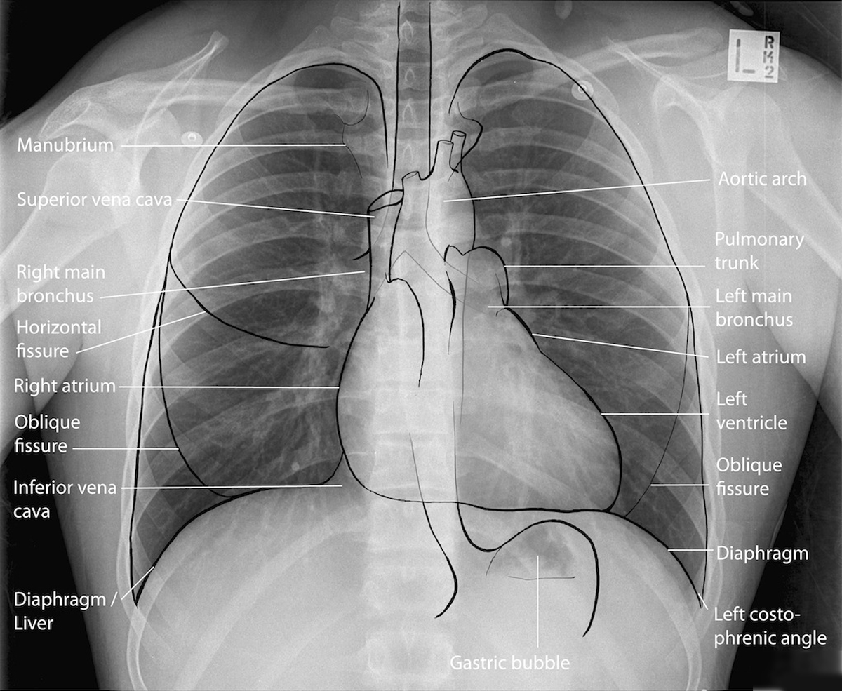
Chest xrays
Chest xrays (CXR) provide a good overview of the state of the airways and airspaces in the lungs. They are useful to check that the lungs are looking as expected and for this reason they are carried out once per year at annual review. They can pick up some particularly problems such as ABPA and when areas of the lung are collapsed, and so they might also be done during chest exacerbations. The resolution of CXRs is relatively low, so a normal-looking CXR does not mean that there is no lung disease.
 | 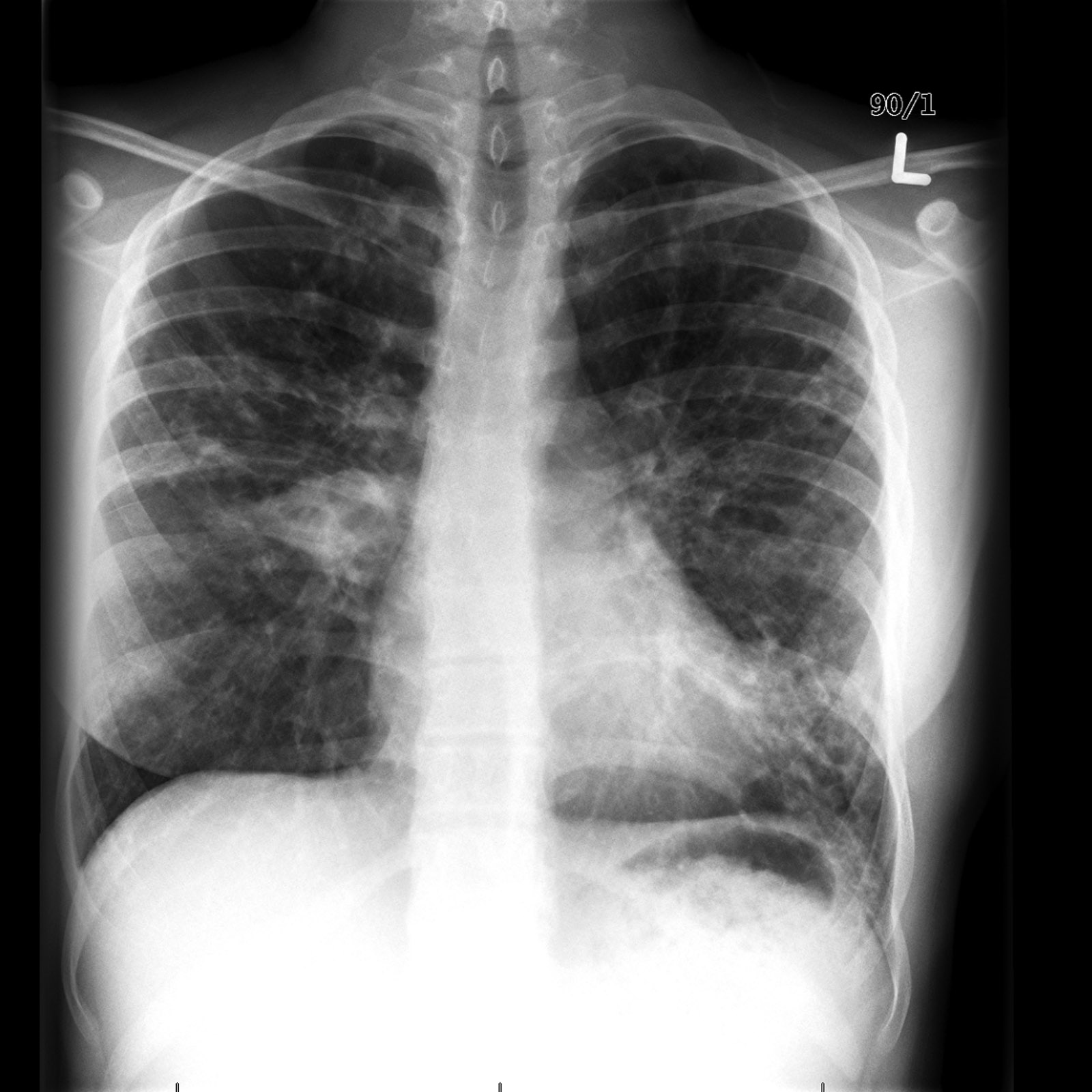 |
| A normal chest xray | A chest xray from a teenager showing moderate changes of CF |
CT scans
Computerised tomography (CT) scans give a much more detailed view of the lungs and airways. CT images provide a view of 'slices' of the lung from top to bottom. They can pick up changes not seen on CXRs. We recommend CT scans are carried out when lung function is poor despite treatment with antibiotics or when lung function is poor without much in the way of cough. The CT scan can indicate whether there are changes consistent with infection. We sometimes use the CT scan to direct us to the best part of the lung to collect airway samples from during bronchoscopy. CT scans cannot be done frequently because of the relatively high radiation dose involved.
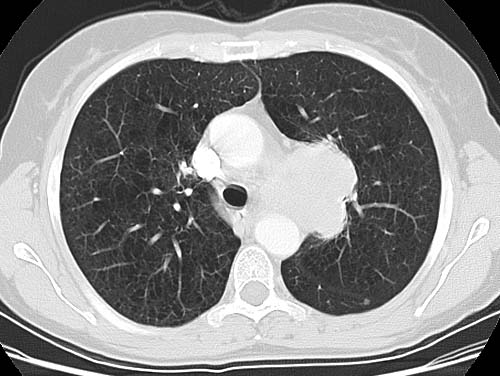 | 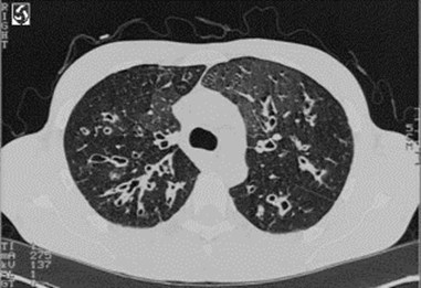 |
| A normal chest CT scan | A chest CT scan from a teenager showing moderate changes of CF |
|